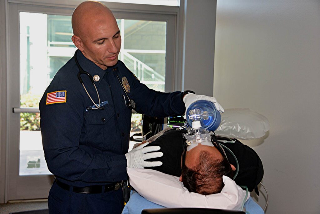
Photo/Rick McClure
When there is a discussion about acute respiratory distress syndrome (ARDS) it is about ventilator management strategies after the patient has been intubated. In fact, the majority of the studies that we use to inform contemporary ventilator management strategies is centered on this population of ARDS patients.
But what about when ARDS develops before intubation and mechanical ventilation? What do these patients look like in the living room or the nursing facility bedroom? How can we manage it?
It can be easy to miss developing or mild ARDS in the prehospital setting. We operate in a resource constrained environment that does not lend itself to prolonged investigations of respiratory complaints outside of the hospital. While we could simply treat them symptomatically, we may miss an opportunity to be more invasive to a greater effect.
As with anything in emergency and critical care, the earlier the problem is identified and treatment initiated, the better things tend to go for the patient. If we can put enough pieces of the story together with what we assess clinically we may even be able to prevent the worst of it.
ARDS Review
ARDS is a pathologic disease process that primarily affects the lungs, however in more advanced stages it contributes to multiple organ dysfunction syndrome (MODS). Through one of several different causes the patient suffers from a combination of non-cardiogenic pulmonary edema and progressively worsening lung compliance. This primarily leads to a hypoxic respiratory failure and then a hypercapnic respiratory failure as the lungs move less and less minute volume.1
There are a few different taxonomies of causes of ARDS, and there have been several different studies that have tried to create sub-categories or phenotypes of ARDS from which clinicians can tailor their treatments.2-3 The idea being that tailored treatment would be the most beneficial thing for the patient.
At the time of this writing, there is not a strong consensus on phenotype titles or if there is a demonstrable benefit to treating ARDS based on phenotype. Some of the different phenotypes of ARDS are: direct vs. indirect; inflammatory vs. non-inflammatory; or pulmonary vs. extrapulmonary.2-4
More commonly, we know the causes of ARDS to be problems like:2-4
- Pneumonia
- Blunt/Penetrating Trauma
- Acute pancreatitis
- Near drowning
- Smoke inhalation
- Shock
- Transfusion-Related Lung Injury (TRALI)
The cause notwithstanding, the subsequent pathophysiologic process of ARDS is relatively predictable, but the damage it causes to the lungs is heterogenous from patient to patient. Cell injury signals the release of pro-inflammatory cytokines and inflammatory mediators into the blood stream.1,4-7
These substances, while performing their primary function of attacking pathogens, damage and destroy the endothelium and glycocalyx in the lungs. This damages the alveolar-capillary membrane and increases its permeability, allowing for fluid to leak into and occupy space in the alveoli.4-7
The fluid that leaks into the alveoli is loaded with pro-inflammatory substances and has a positive oncotic pressure, which means that it continually draws more and more fluid to it. This contributes to a persistent worsening of the edema. 4-7
The compliance problem is due to the edema and the loss of surfactant. The alveoli are “stiff” and atelectatic, making it difficult to expand them and keep them expanded during inspiration.4-7
The problem is that it causes the patient (or a ventilator) to exert more force to move air into the alveoli. As they lose surfactant, the alveoli collapses which reduces the overall surface area in the lungs for ventilation and oxygenation.
These two issues lead to hypoxic respiratory failure. The edema inhibits the diffusion of oxygen from the alveoli into the blood stream, and the poor compliance leads to less minute ventilation moving into and out of the lungs.1,4-7
What you are left to deal with is hypoxia and shunt physiology. With this type of right-to-left shunt, the patient will not respond to simple increases in FiO2, requiring more invasive and potentially injurious treatments (if not administered carefully) to restore the patient’s ability to oxygenate.
Don’t Be SILI…
How can this devastating damage be caused without the use of a mechanical ventilator or other form of positive pressure ventilation? A patient in respiratory distress has a very intense respiratory drive, effort, and abnormal respiratory patterns.8
This combination of respiratory mechanics can stress the lung tissues and generate an inflammatory response that leads to ARDS. This is more likely to happen when there is already a disease process present that is influencing the lung mechanics. The stronger the respiratory drive the faster onset of this self-induced lung injury (SILI or P-SILI).1,8
If You Are Not Early, You Are Late
As with anything in emergency or critical care, the earlier ARDS is identified the easier it is to manage. In fact, if done well, its progression to MODS and death can be prevented in some cases (this is very heavily dependent on the underlying cause and the patient’s medical history).
How can it be identified in the resource constrained prehospital environment? There are a number of options that can support a suspicion of ARDS, but this environment typically lacks the tools needed to definitively meet the diagnostic criteria for ARDS.
Tools to Identify It
SpO2/FiO2 ratio: Not to be confused with the PaO2/FiO2 ratio that is typically used to gauge the presence and severity of ARDS. The SpO2/FiO2 ratio is something that can be used when there is no way to obtain an ABG, as is often the case in the prehospital and transport settings. This ratio is obtained by dividing the SpO2 by the FiO2 with a value <315 being indicative of hypoxemia consistent with ARDS.9-10 This method can only be used when SpO2 is <97% due to the influence of the shape of the oxyhemoglobin dissociation curve.9-10
Lung Ultrasound: LUS to identify ARDS is an emerging area of research. The Kigali modification to the Berlin definition of ARDS suggests the use of bilateral lung ultrasound to be included in the diagnostic criteria for ARDS. This modification has some important implications for use in resource constrained clinical settings.11-12
Clinicians typically use ultrasound to identify several thoracic abnormalities like pneumothorax, effusions, and pneumonia.11-12 LUS specifically for identifying ARDS does lack evidentiary support as of this writing. Studies in its use with ARDS show high sensitivity, but low specificity with LUS.11-12 This is evident by the purported “positive” findings for ARDS on LUS are the presence of B-lines or consolidation in at least one area on both sides of the chest.11-12 But these findings are not terribly specific to ARDS. Implementing LUS in this manner seems straightforward and it will be interesting to see what further research yields in this area.
How Can It Be Managed?
Higher Fio2 may or may not do it.
ARDS is primarily an oxygen diffusion problem. The alveoli are filled with fluid that causes an increase in the time it takes to diffuse oxygen through the alveolar capillary membrane and into the blood stream.
This creates shunt physiology, and in this particular flavor of it, there are areas of the lungs that are receiving blood flow but no gas exchange. Simply increasing the FiO2 with a non-rebreather mask that will deliver 60%-90% FiO2 may not work.
The problem, as previously mentioned, is the edema in the alveoli, and oxygen does not diffuse well through fluid. In the acute phase there is no way to redistribute the fluid out of the alveoli and facilitate better oxygen diffusion. This means that we have to maximize the form and function of the healthy lung units (known colloquially as “baby lung”).
NIV shows some promise.
Non-Invasive Ventilation (NIV) for ARDS has spotty support in the literature. Some studies show that it is wise to avoid it since a failed attempt at NIV will result in a worse disease burden, but there is some disagreement about whether the worsening disease burden is directly related to NIV failing to correct the hypoxemia or if NIV failure is more of an indicator of severe underlying disease.13 It is still quite indicated for hypoxemic respiratory failure and pulmonary edema, the two physiologic hallmarks of ARDS.
To maximize the effect of the additional FiO2, the addition of positive pressure in the form of PEEP will be essential. Functional residual capacity can be reduced by as much as 30% in ARDS, and PEEP, delivered either by CPAP or BiPAP, can recruit and stent open the collapsed alveoli.5, 13-15
This can help overcome the shunt physiology and will also be critical for protecting the healthy “baby-lung” from damage and inflammation. The term “baby-lung” is used to describe the areas of the lungs that are still functioning and aerating normally. These areas are small and fragile, like a baby, hence the term “baby-lungs.”
The NIV strategy has limitations. PEEP can recruit collapsed alveoli, but only the lung units that are recruitable.13-15 For much of the lung surface area it is damaged and inoperable until the patient has had time to heal. PEEP and NIV may not reverse ARDS, but maybe it can stop it from getting any worse.
To Close Out
ARDS is not a problem specially reserved for those patients receiving mechanical ventilatory support. Prehospital teams need to maintain a high index of suspicion for ARDS in patients with common complaints such as pneumonia and pancreatitis (just to name a few).
It is important to get aggressive treatments in place to protect the healthy lung area and maintain adequate oxygenation.
References
1. Swenson KE, Swenson ER. Pathophysiology of Acute Respiratory Distress Syndrome and COVID-19 Lung Injury. Crit Care Clin. 2021 Oct;37(4):749-776. doi: 10.1016/j.ccc.2021.05.003. Epub 2021 May 28. PMID: 34548132; PMCID: PMC8162817.
2. Reilly JP, Calfee CS, Christie JD. Acute Respiratory Distress Syndrome Phenotypes. Semin Respir Crit Care Med. 2019 Feb;40(1):19-30. doi: 10.1055/s-0039-1684049. Epub 2019 May 6. PMID: 31060085; PMCID: PMC6657496.
3. Sinha P, Calfee CS. Phenotypes in acute respiratory distress syndrome: moving towards precision medicine. Curr Opin Crit Care. 2019 Feb;25(1):12-20. doi: 10.1097/MCC.0000000000000571. PMID: 30531367; PMCID: PMC6814152.
4. Matuschak GM, Lechner AJ. Acute lung injury and the acute respiratory distress syndrome: pathophysiology and treatment. Mo Med. 2010 Jul-Aug;107(4):252-8. PMID: 20806836; PMCID: PMC6188356.
5. Biuzzi C, Modica E, De Filippis N, et al. Old and New Definitions of Acute Respiratory Distress Syndrome (ARDS): An Overview of Practical Considerations and Clinical Implications. Diagnostics. 2025; 15(15):1930. https://doi.org/10.3390/diagnostics15151930
6. Luo J, Yu H, Hu YH et al. Early identification of patients at risk for acute respiratory distress syndrome among severe pneumonia: a retrospective cohort study. J Thorac Dis. 2017 Oct;9(10):3979-3995. doi: 10.21037/jtd.2017.09.20. PMID: 29268409; PMCID: PMC5723858.
7. Deshwal H, Elkhapery A, Ramanathan R et al. Patient-Self Inflicted Lung Injury (P-SILI): An Insight into the Pathophysiology of Lung Injury and Management. J Clin Med. 2025 Feb 27;14(5):1632. doi: 10.3390/jcm14051632. PMID: 40095610; PMCID: PMC11900086.
8. Sklienka P, Frelich M, Burša F. Patient Self-Inflicted Lung Injury-A Narrative Review of Pathophysiology, Early Recognition, and Management Options. J Pers Med. 2023 Mar 28;13(4):593. doi: 10.3390/jpm13040593. PMID: 37108979; PMCID: PMC10146629
9. Erlebach R, Pale U, Beck T, Markovic S, Seric M, David S, Keller E. Limitations of SpO2 / FiO2-ratio for classification and monitoring of acute respiratory distress syndrome-an observational cohort study. Crit Care. 2025 Feb 19;29(1):82. doi: 10.1186/s13054-025-05317-7. PMID: 39972458; PMCID: PMC11837723.
10. Matthay MA, Arabi Y, Arroliga AC, et al. A New Global Definition of Acute Respiratory Distress Syndrome. Am J Respir Crit Care Med. 2024 Jan 1;209(1):37-47. doi: 10.1164/rccm.202303-0558WS. PMID: 37487152; PMCID: PMC10870872.
11. Lazzeri C, Peris A. The Kigali modification of the berlin definition: a new epidemiological tool for ARDS? J Thorac Dis. 2016 Jun;8(6):E443-5. doi: 10.21037/jtd.2016.03.84. PMID: 27294247; PMCID: PMC4886026
12. Smit MR, Hagens LA, Heijnen NFL, et al. DARTS Consortium members. Lung Ultrasound Prediction Model for Acute Respiratory Distress Syndrome: A Multicenter Prospective Observational Study. Am J Respir Crit Care Med. 2023 Jun 15;207(12):1591-1601. doi: 10.1164/rccm.202210-1882OC. PMID: 36790377; PMCID: PMC10273105.
13. Tucci MR, Costa EL, Nakamura MA, Morais CC. Noninvasive ventilation for acute respiratory distress syndrome: the importance of ventilator settings. J Thorac Dis. 2016 Sep;8(9):E982-E986. doi: 10.21037/jtd.2016.09.29. PMID: 27747041; PMCID: PMC5059316.
14. Bellani G, Laffey JG, Pham T, et al. LUNG SAFE Investigators; ESICM Trials Group. Noninvasive Ventilation of Patients with Acute Respiratory Distress Syndrome. Insights from the LUNG SAFE Study. Am J Respir Crit Care Med. 2017 Jan 1;195(1):67-77. doi: 10.1164/rccm.201606-1306OC. PMID: 27753501.
15. Dunand A, Beysard N, Maudet L, et al. Management of respiratory distress following prehospital implementation of noninvasive ventilation in a physician-staffed emergency medical service: a single-center retrospective study. Scand J Trauma Resusc Emerg Med. 2021 Jun 29;29(1):85. doi: 10.1186/s13049-021-00900-7. PMID: 34187538; PMCID: PMC8240431.

Cody Winniford is a flight paramedic and base manager in Baltimore, MD. He has a passion for sharing his professional experience in EMS and management. Cody’s clinical and leadership development background spans both military and civilian settings and has served in several capacities as a leader and prehospital clinician. He specializes in air medical and critical care transport, as well as organizational development and leadership development. He is an active speaker on various leadership and clinical topics and is an established and successful educator for prehospital clinicians of all levels. He has a passion for human performance improvement and the mental health and performance aspects of prehospital care.