Shutterstock/Your Hand Please
With rare exceptions, the emergency vehicles that we drive run using internal combustion. While gasoline and diesel engines operate based on different principles, they both combine fuel with air to produce energy that is harnessed for power, while also emitting exhaust gases as a byproduct.
One measure that helps to monitor engine output is a tachometer, which displays revolutions per minute (RPMs). In a similar manner, our bodies create energy by combining fuel, in the form of food, and oxygen from the air that we breathe.
Through the processes of respiration, perfusion and metabolism (RPM), energy in the form of adenosine triphosphate (ATP) is created that enables the functioning of body systems, while byproducts are produced to be eliminated. One way to measure the efficiency of the process is the use of capnography.
Carbon Dioxide Generation
Human bodies require both pulmonary and cellular respiration. We inhale oxygen into our lungs, which diffuses into the capillaries that surround alveoli. Diffusion occurs from an area of higher pressure to a lower one.
Since the venous blood that is pumped from the right ventricle is somewhat oxygen-depleted, the freshly inhaled oxygen flows into the capillaries for the return trip to the left atrium. Oxygenated blood is pumped throughout the body from the left ventricle into the arterial system.
Digestion of the food that we eat, primarily carbohydrates, produces glucose, which enters cells with the assistance of insulin. Glycolysis breaks down glucose into pyruvic acid. With good perfusion, and the presence of adequate oxygen, aerobic metabolism will occur, and energy in the form of ATP is produced, with byproducts of carbon dioxide (CO2) and water (H2O) that diffuse back into the bloodstream.
This movement of gases in and out of the cells comprises cellular respiration.
If there is inadequate oxygen (hypoxia) or inadequate perfusion to the cells, as with shock, the pyruvic acid will ferment into lactate, also known as lactic acid. This inefficient process is called anaerobic metabolism which, if continued, results in lactic acidosis. Very little energy is produced along with reduced CO2 and H2O byproducts.
With normal functioning, a healthy body will transport metabolic waste to the kidneys, where excess water is removed, and the lungs, where CO2 is exhaled. Since normal air should not contain CO2, the higher pressure in the return venous blood diffuses into the alveoli for exhalation. This exhaust gas can be measured with capnography.
Development of Capnography in EMS
Early capnography was generally limited to use within healthcare facilities, particularly in the world of anesthesiology. CO2 detection in the prehospital arena came about through the introduction of colorimetric devices that indicated the presence of the gas by color change and was essentially used to confirm endotracheal intubation.
Lack of color change indicated improper tube placement or death. If the patient had adequate perfusion, the change was pronounced. In low-flow states, such as cardiac arrest with CPR, change was more subtle. The devices merely indicated a limited or adequate amount of CO2, as opposed to none.
Around the turn of the century, capnography was added as monitors used in the field became increasingly sophisticated. It was primarily used for tube confirmation but was also able to provide a quantitative analysis of the amount produced.
Through the adoption of advancing technology, along with the availability of devices that incorporate the sensor into a nasal cannula, its use has become not only commonplace but recommended for many conditions.
Providers have learned not only the quantitative analysis of the amount of detected CO2, but also the qualitative analysis of the waveform.
A Measure of Metabolism and Ventilation
From a quantitative perspective, the amount of CO2 detected is reflective of both metabolism and ventilation. With good readings, a properly inserted connector that eliminates sample dilution, and under normal physiological conditions, end-tidal readings should be in the 35-45 mmHg range.
A hyperventilating patient will have low readings, but the cause must be determined and might vary from mere anxiety to compensation for a metabolic condition, such as acidosis.
High readings might be indicative of inadequate ventilation, such as with medication-induced respiratory depression, or preexisting conditions such as COPD.
Apart from respiratory conditions, remember that CO2 is a byproduct of metabolism. Conditions such as sepsis can lead to reduced metabolism and an increase in respiratory rate to compensate for developing acidosis, resulting in low readings.
During shock, perfusion is reduced and therefore so is metabolism and CO2 production.
Cardiac arrest leads to no metabolism other than what is facilitated by CPR. Since CPR produces a fraction of normal blood flow, CO2 will be relatively low in the absence of ROSC, which may be indicated by a sudden rise.
Prolonged asystole with persistently low capnography readings despite properly performed CPR is an indication of death.
Capnography Waveforms
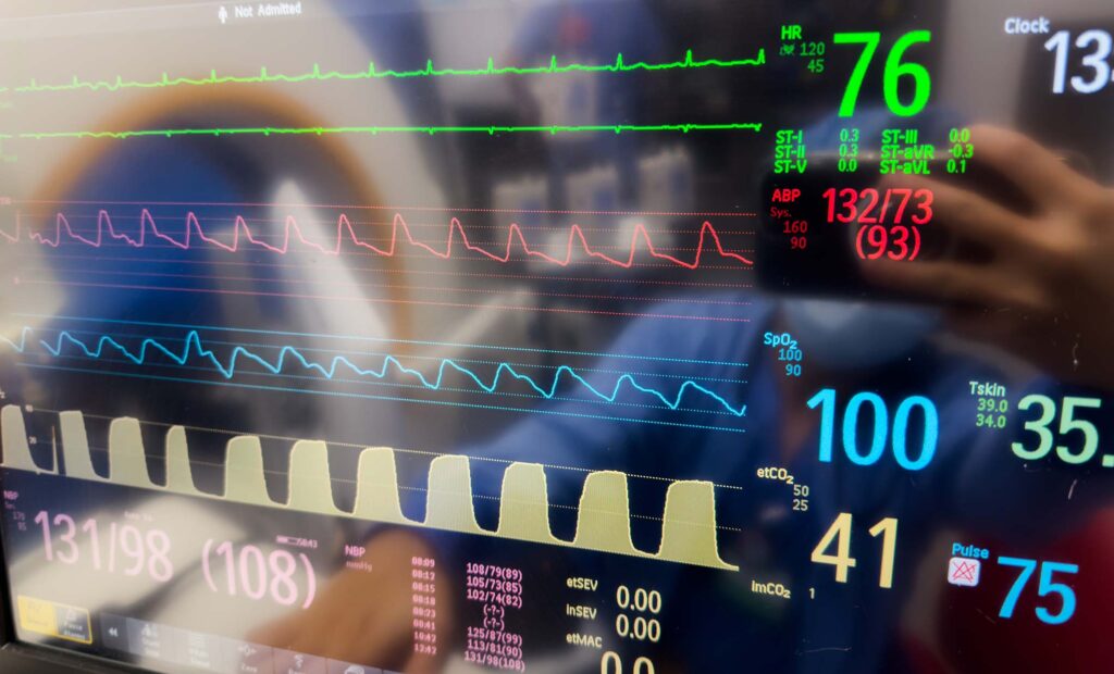
A normal capnograph will have a baseline between breaths, an almost vertical rise if airways are not restricted, a slightly rising plateau with a peak that provides our numerical reading, and then a sudden fall during inhalation or positive pressure ventilation.
Each waveform represents one breath and a sudden loss in an unresponsive patient is indicative of respiratory arrest.
Tachypnea results in more waveforms on the screen and lower levels of end-tidal CO2. Inadequate ventilation, such as is unfortunately seen with all-too-common narcotic overdoses, results in fewer waveforms and a higher level.
One of the common abnormal patterns has a shark fin shape and is indicative of bronchoconstriction. Conditions such as asthma are air trapping as exhalation is restricted.
Since the CO2 does not flow as rapidly through the restriction, the upstroke becomes arced. This pattern can assist in determining the underlying condition of the patient.
As an example, wheezes might be heard early with congestive heart failure (CHF) but the initial treatment differs from a COPD exacerbation, which generally produces a pronounced arc.
It can also reflect the efficacy of care; relief of bronchoconstriction will be displayed with a more upright pattern, while worsening of the arc might indicate that more advanced treatment, such as intramuscular epinephrine, is required.
There are other abnormal patterns that should be learned over time, from more than one article, to improve diagnostic capability.
Much like the vehicles that we drive, our bodies depend on a sufficient mixture and transport of fuel and air. With either, adequate energy production results in exhaust gases; capnography is a means for measuring human exhaust.
Vehicles and humans both require precise conditions that result in proper operation. Vehicle engine output can be measured in RPMs; human energy production is also based on RPMs but, in the body, they are respiration, perfusion and metabolism and are reflected in capnography.
In either case, normal function is dependent upon having adequate RPMs.
Lew has been involved in EMS since 1971, when he was initially certified as an EMT, and is also a former fire chief. He spent several of those years, often simultaneously, in both prehospital and hospital-based care, and remains active in paramedic training.

