
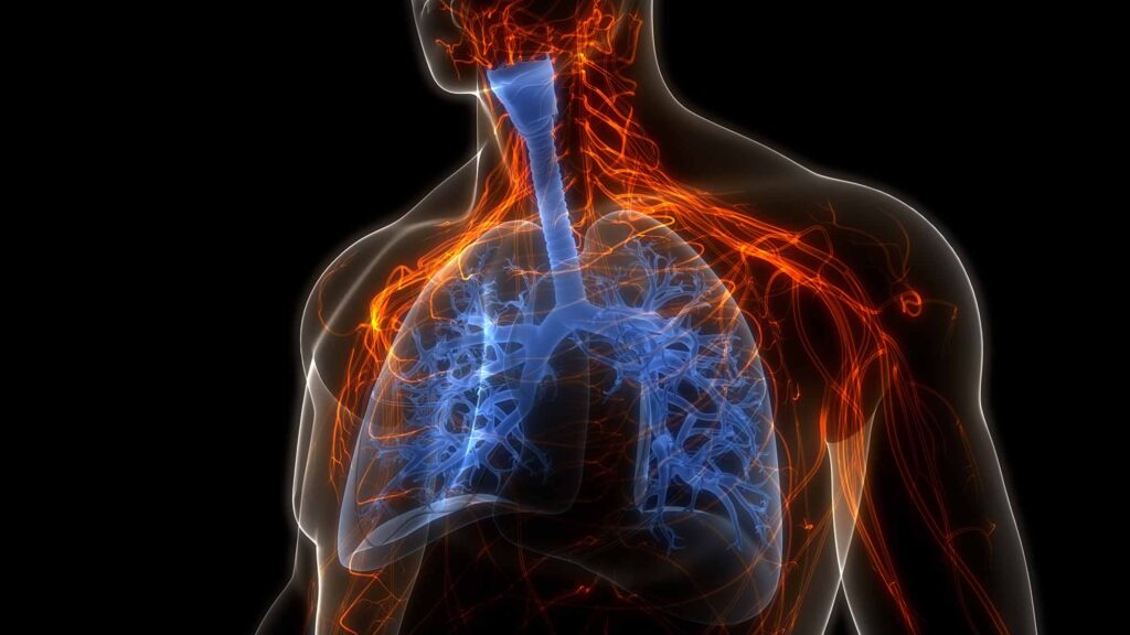
3D illustration of the human respiratory system. (Shutterstock/Magic mine)
There are aspects of vent management with the obstructive lung disease patient population that is simply noise. What I mean by “noise” is that there are things we should expect to see that are simply sequelae of the disease, not things that we fix directly, and won’t be fixed until we fix the problem that causes those sequelae. The noise is the predictable consequence(s) that we know is going to be there because the patient has been suffering an air flow pattern restriction. You could think of this as “physiologic background noise.”
On the other hand, there are signals that the patient is not responding to or tolerating therapies and their condition is getting dangerously worse. Anything that signals worsening hyperinflation or Auto-PEEP is something we absolutely have to pay attention to and work diligently to correct.
Depending on the type of ventilator you use, you can have a vast array of signals and noise coming at you in the course of a transport, and even the most well-meaning clinician can find themselves in trouble by chasing the noise. A “knobologist” is likely to perform maneuvers that “fix” high CO2 or ETCo2 can absolutely make the patient’s situation more dire by worsening air flow and ventilation.
Part 2 of this series (See: Signals and Noise: Vents and Obstructive Lung Disease – Part 1) is about what to pay attention to versus what can wait or be ignored when ventilating the obstructive lung disease patient. Sometimes, it is as simple as changing an alarm parameter to quiet the noise, and other times you are receiving a signal that there is a serious problem to address that requires an advanced maneuver and a little patience to correct.
Expiratory Flow Returning to Baseline – Signal1-6
If the expiratory flow waveform is not returning to baseline, there is a risk that the patient is air trapping, which is dangerous to the patient for a couple of different reasons. First, it increases the resistive forces making it more difficult for air to move into the alveoli. Next, it compresses the pulmonary vasculature (and the inferior vena cava in some cases) creating conditions favorable for hemodynamic collapse.
The compression of these vessels increases the pulmonary artery pressure and can increase the pressure that the right ventricle has to pump against. This obstructive physiology leads to RV failure and subsequently the rest of the cardiovascular system can fail. To confirm, look for the total PEEP (if your ventilator has this setting).
- Total PEEP > set PEEP = air trapping (in the setting of obstructive lung disease).
- Air trapping + high peak pressures + hypotension (not attributed to another disease process) = obstructive shock physiology until proven otherwise.
Pplat increasing – Signal3,7-9
The Pplat is the guardrail for all of your maneuvers. With these patients, we expect high peak pressure. Their entire pulmonary system is under incredible pressure that will invariably make your ventilator alarm over and over and over, but again, its expected due to the nature of the obstructive pathology.
A high Pplat, or plateau pressure, is not expected. Pplat is a measurement of how much pressure is in the alveoli, and we know that when it is less than 30 the patient is not in danger of lung injury. However, when it is greater than 30, we know that the alveoli are distended and that damage is occurring with each breath.
This damage and ensuing inflammation leads to the development of ARDS, dramatically complicating this patient’s clinical course. Barotrauma is another risk to be aware of when the pPLAT is above 30, and particularly so in this patient population.
Hypercapnia – Noise2, 5-10
Hypercapnia in this case is “noise.” By “noise” I mean that it is something that is an expected sequelae of the pathology you are dealing with, not a problem to be attacked in isolation. These patients have expiratory flow limitation which by definition means that they are retaining CO2 due to an inability to exhale it.
The astute clinician will know that they cannot “fix” hypercapnia by increasing the respiratory rate for these patients (and that doing so will likely signal a worsening not an improvement in air flow), lest they steal from the exhalation time. They will also remember that a state of permissive hypercapnia is a part of managing these patients. That hypercapnia is allowed to persist until the patient’s pH falls below 7.2, at which point the strategy needs to shift.
This is by no means a recommendation to “ignore” hypercapnia. Rather it is an important perspective to have when managing these patients so that you do not get dragged down into the weeds and lose awareness of the airflow problem that is creating the hypercapnia. They will experience cardiac arrest from auto-PEEP before they suffer any sort of devastating effects from hypercapnia.
Ppeak – Noise8
As previously stated, we expect high peak pressures with these patients. We expect this because of the combination of narrow airways and the trapped air in the alveoli causing the ventilator to use more pressure to move the volume through the narrow airways and into the alveoli.
Chasing the Ppeak or PIP (depending on your vent), is perhaps the most common cognitive trap to fall into. It is a reflection of airway resistance, not the pressure in the alveoli. The reflex of lowering tidal volume to reduce the Ppeak will likely leave your patient being under ventilated and worsen their condition.
Things only get worse if we end up fighting with the ventilator. The Hamilton T-1 (my favorite) protects the patient from over pressuring the lungs by stopping breath delivery when the pressure rises to within 10cmH2O of the pressure limit. Meaning, the patient’s breaths are being cut off.
To overcome this clinicians may need to increase these pressure limits beyond the anticipated PIP/Ppeak. This can only be done with the understanding that the Pplat is the safety net and must be measured frequently in order to ensure that lung injury is not occurring.
If after this maneuver, there are still problems with alarms and flow limitation alerts, you have to work through your standard troubleshooting steps to rule out under sedation/poor pain control, pneumothorax, kinked tube/feedback lines, and mucus plugging at the end of the tube.
Take Home Points
Treating a mechanically ventilated obstructive lung disease patient requires a quiet mind in a loud an chaotic environment. Knowing what you need to pay attention to can make things just a little quieter for you.
The deeper your understanding of the disease and what to expect the patient to be dealing with makes it easier to look past an alarm that is begging for your attention, but robbing you of the focus on signals of badness.
Trust your strategy of lower respiratory rates and tidal volumes of 8cc/kg IBW that we talked about in Part 1.
References
1. Stather DR, Stewart TE. Clinical review: Mechanical ventilation in severe asthma. Crit Care. 2005;9(6):581-587. doi:10.1186/cc3733
2. Gayen PK. Mechanical ventilation in obstructive airway diseases. In: Principles and Practice of Mechanical Ventilation. IntechOpen; 2019.
3. Abid S, Nava S. Ventilatory strategies in obstructive lung disease. Clin Chest Med. 2006;27(3):489-501. doi:10.1016/j.ccm.2006.04.004
4. Weingart S. EMCrit 15 – The severe asthmatic. EMCrit Blog. Published December 8, 2009. Accessed October 29, 2025. https://emcrit.org/emcrit/severe-asthmatic/
5. Ahmed SM, Athar M. Mechanical ventilation in patients with chronic obstructive pulmonary disease and bronchial asthma. Indian J Anaesth. 2015;59(9):589-598. doi:10.4103/0019-5049.165856
6. Jubran A. Setting positive end-expiratory pressure in the severely obstructive patient. Curr Opin Crit Care. 2024;30(1):89-96. doi:10.1097/MCC.0000000000001131
7. Demoule A, Brochard L, Dres M, et al. How to ventilate obstructive and asthmatic patients. Intensive Care Med. 2020;46(12):2436-2449. doi:10.1007/s00134-020-06291-0
8. Lodeserto F. Simplifying mechanical ventilation – Part 4: Obstructive physiology. REBEL EM. Published June 22, 2018. Accessed August 31, 2026. https://rebelem.com/simplifying-mechanical-ventilation-part-4-obstructive-physiology/
9. Zaidi SF, Shaikh A, Khan DA, Surani S, Ratnani I. Driving pressure in mechanical ventilation: A review. World J Crit Care Med. 2024;13(1):88385. doi:10.5492/wjccm.v13.i1.88385
10. Farkas J. PulmCrit: The crashing asthmatic. EMCrit Blog. Published 2014. Accessed August 31, 2026. https://emcrit.org/pulmcrit/crashing-asthmatic/

Cody Winniford is a flight paramedic and base manager in Baltimore, MD. He has a passion for sharing his professional experience in EMS and management. Cody’s clinical and leadership development background spans both military and civilian settings and has served in several capacities as a leader and prehospital clinician. He specializes in air medical and critical care transport, as well as organizational development and leadership development. He is an active speaker on various leadership and clinical topics and is an established and successful educator for prehospital clinicians of all levels. He has a passion for human performance improvement and the mental health and performance aspects of prehospital care.
